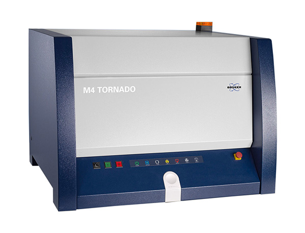


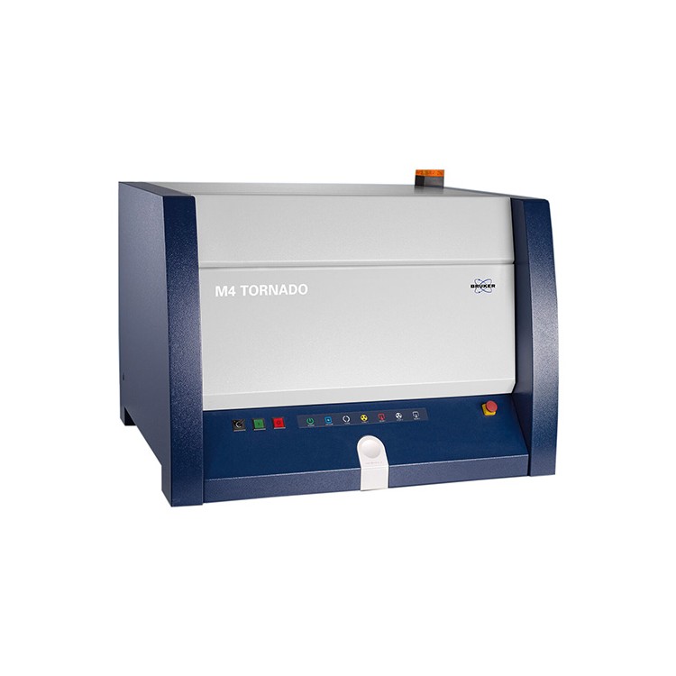


M4 Tornado Micro-XRF Spectrometer

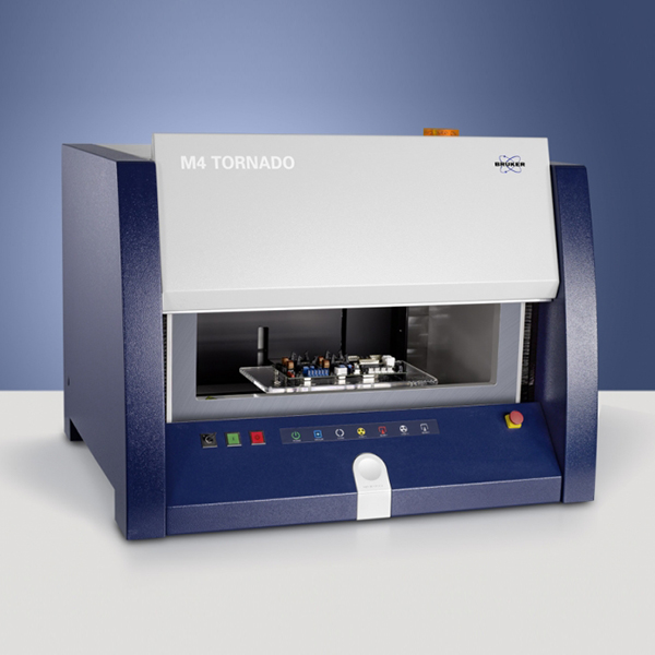
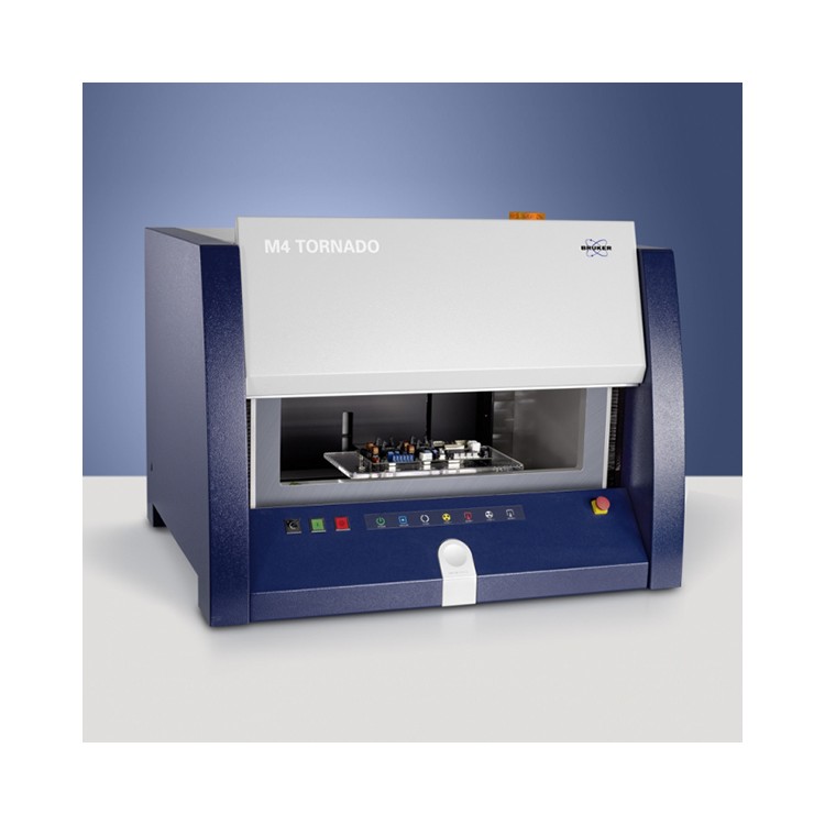
The Bruker M4 Tornado Micro-XRF Analyzer is suitable for high-sensitivity, non-destructive elemental analysis (Na11-U92) of large, uneven, irregular, and even small samples and packages. It is a new tool for research in sample composition analysis and distribution patterns, offering scholars new perspectives from macro, micro, and meso-microscopic angles.
Employing a multi-mode capillary focusing lens to concentrate the excitation light into an extremely small area (<20um) for superior spatial resolution and element imaging analysis. Different types of samples can be analyzed directly through simple sample preparation, or even without preparation. The equipment outputs high-precision element distribution maps with a resolution as high as 40 megapixels, 7168*5582 pixels.

Application Cases
1. Plant Environment Applications
The micro-area X-ray fluorescence spectrometer equipment can rapidly perform non-destructive scanning on various types of animal and plant samples, allowing for timely observation of the migration process of elements in the roots, stems, leaves, flowers, and seeds of plant samples, as well as changes in element content at different stages and element imaging analysis. It can also simultaneously conduct soil heavy metal pollution monitoring, plant nutrition and stress resistance research, insect morphology studies, and biological tissue compatibility research.
Utilizing micro-XRF analysis on rice leafs, we identified several SR proteins as key regulators of phosphorus nutrition by studying the translocation and enrichment patterns of P in wild-type and multiple mutant lines, and demonstrated that three SR protein-coding genes regulate the absorption and inter-stem/leaf migration of phosphorus in rice.

2. Technological Archaeology
In archaeological research, X-ray fluorescence spectrometry is primarily used to determine the composition of ancient artifacts, thereby inferring and judging the social and cultural aspects of the time. Micro-area X-ray fluorescence spectrometry not only provides quantitative data but also visually presents the distribution of components. It is mainly applied to the compositional analysis of cultural relics such as ceramics, bronze objects, gold and silverware, ancient buildings, painting pigments, and ancient architectural structures.
The micro-XRF analysis of Neolithic white pottery reveals distinct layers on the interior wall of the pottery samples. The distribution of Ca elements indicates that these enriched regions correspond precisely to the locations of the layers on the inner wall, suggesting that this residue is a calcareous type of material rich in calcium. This discovery illustrates that white pottery had already been used as a practical utensil during the Neolithic era.

3. Earth Sciences field
The micro-area X-ray fluorescence spectrometer is a crucial analytical instrument in geochemistry, primarily used for non-destructive, high-speed scanning and elemental distribution imaging of samples such as rocks, meteorites, and fossils, as well as for mineral identification, quantification, and classification.
By utilizing micro-area XRF analysis on geological thin sections or ore roughs, the elemental distribution pattern on the surface of the thin sections can be obtained. Additionally, by using professional software, information on mineral distribution can be acquired, thereby obtaining the macroscopic regularities of the sample's composition distribution.

4. Material Science
Applied to research on film coating thickness and uniformity; imaging of component and distribution of metal particles in nanometer films; identification of ionic corrosion on metal surfaces and distribution of coating process elements; characterization of alloy sample composition gradients; quality control and fault detection of electronic components such as detector chips, as well as quantitative identification and failure analysis of contact points and welding joints.
Utilizing micro-XRF analysis on concrete provides a clear view of the distribution and penetration depth of Cl elements. This enables identification of harmful elements and their erosion status, facilitates the assessment of material structure, and supports data for structural strength repair.

5. Biotechnology
Applied to biological tissue sections; imaging the penetration and distribution of elements interacting with implanted materials and biological tissue; and analyzing harmful substances in biomimetic materials, etc.
By utilizing micro-XRF analysis on artificial ligaments, the ligaments are retrieved from within the plant organism after a period of time for testing. The distribution maps of Ca and Cl elements reveal the mineral deposits within the artificial ligaments, as well as the presence of any impurity elements or by-products.

6. Criminal Investigation and Public Security
Utilize micro-region XRF analysis for residue analysis of firearm residues on clothing. Most residues are in tiny particles, which cannot be detected directly. An ideal approach is to image the distribution of these particles at high resolution. Imaging with Cu, Pb, and Ba elements can yield data related to the type of sample, distance, and shooting angle, among others.