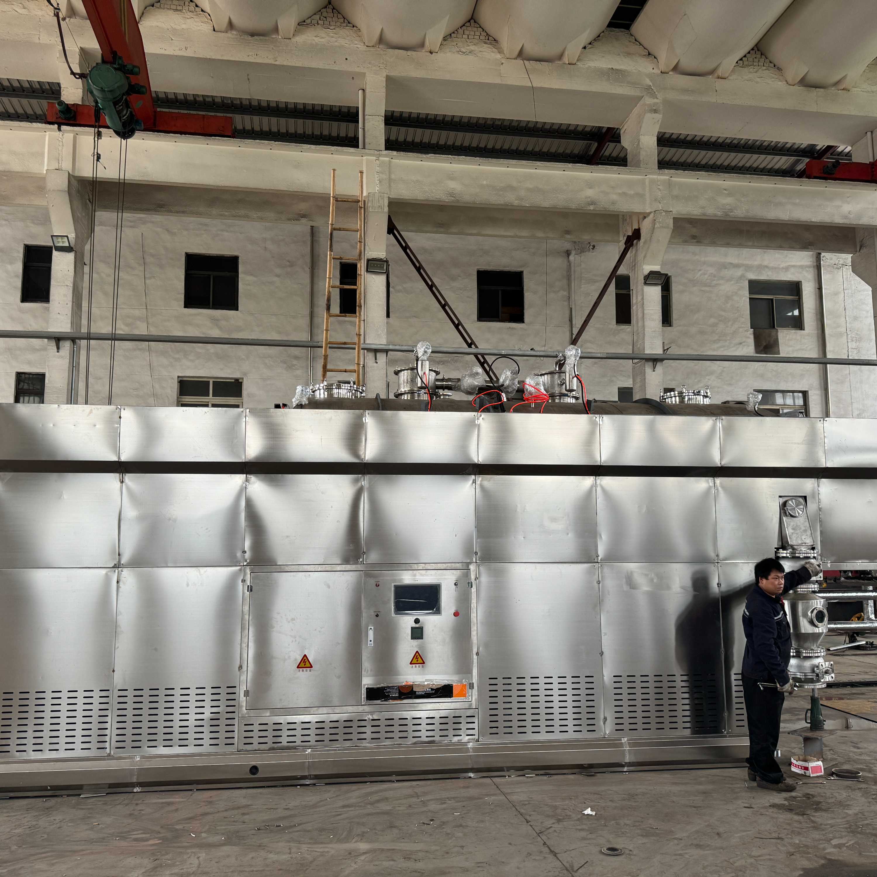


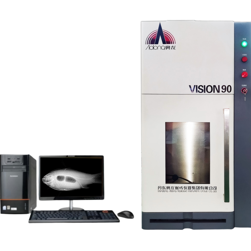


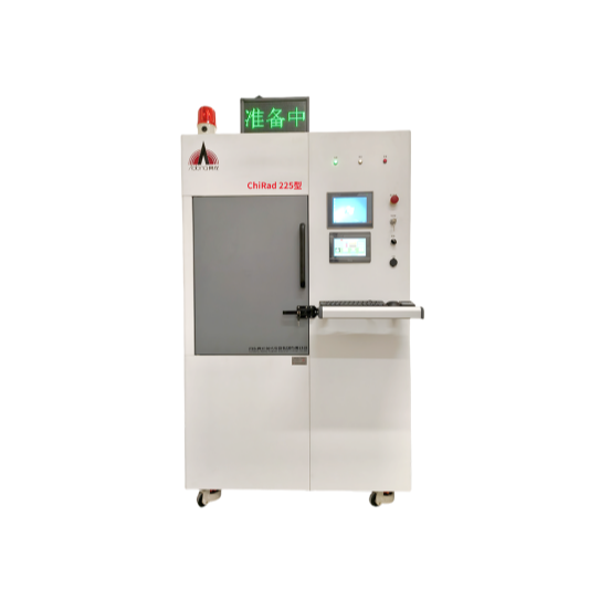
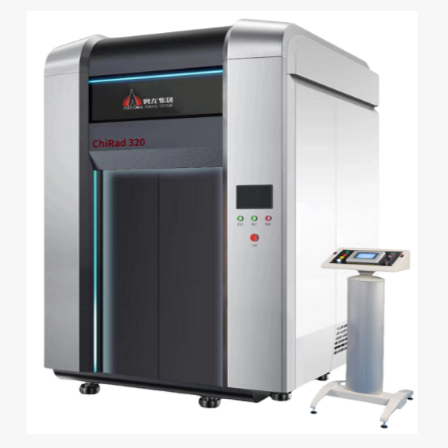


Micrometer-scale X-ray imaging system
Application Fields
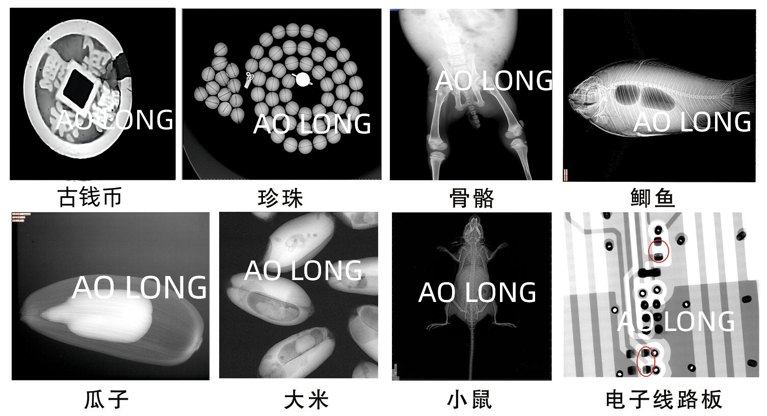
● Live Animal Imaging for Small Animals: Mouse/rabbit research, phenotyping, bone regeneration, tumor growth, osteoporosis/orthopedic research, angiography, pulmonary studies
● Oral Research: Tooth Imaging, Dental Impression, Denture Imaging, Tooth Structure Imaging, Small Animal Limb Contrast Imaging Studies
● Pre-screen for Micro-CT to shorten experimental time
● Seed Imaging: Assess seed viability, contamination, and pest infestation
●Marine Fish Imaging: Fish Skeletons, Bubbles, etc. Research
● Non-destructive Testing: Printed Circuit Boards, Automotive Parts, Aerospace Components, Medical Devices, Electrical/Mechanical Parts, Injection Molds, Castings, Gemstone Inspection
Features
●Easy to use, operators do not require professional X-ray knowledge
● No additional radiation protection setup required
Under full-load operation, the leakage dose of X-rays from the cabinet surface conforms to national standards.
● Configure professional image processing software
● Specimen Size Measurement
● Establish independent storage files to save experimental setup data
● Equipped with shooting control and image processing workstations
● Images can add pseudo-color labels to key areas of the detected image for easier observation.
● Experimental results are savable and printable.
Optional accessories:
● Defect Automatic Identification Feature
3D Imaging Software
Key Technical Parameters
● Voltage: 100KV
● Focal Size: 5μm
● Detector pixel size: 49μm
● Analog-to-Digital Conversion: 16-bit
●Maximum detectable area: 15cm x 10cm
● Geometric Magnification: 20x
●Dimensions: L 60" x W 50" x H 100"
●Weight: 110 kg
Customizable systems available, with configurations to match experimental requirements.
Inspection Case