

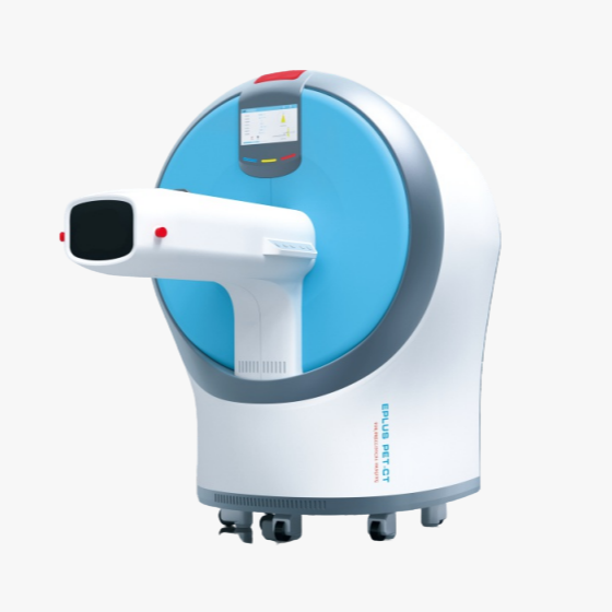


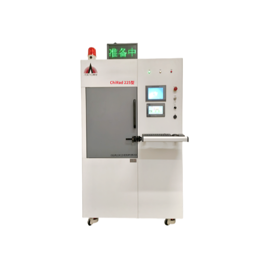


Live Animal PET-CT
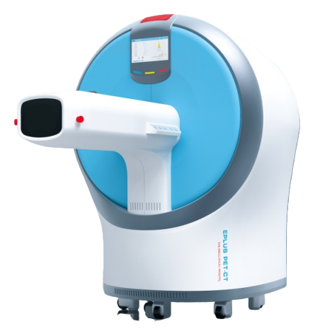
【Core Applications】
Supports preclinical in-vivo PET-CT imaging for a variety of laboratory animals, including mice, rats, rabbits, monkeys, and more, for research on diseases such as tumors, nerves, cardiovascular, and brain, as well as for molecular probe development and drug efficacy evaluation.
【Core Performance Advantages】
- PET axial field of view reaches 200mm (longest in the industry), single bed completes whole-body dynamic imaging of rats
1.5mm detector pixel + SiPM readout technology, achieving sub-millimeter high-definition PET imaging
- Autonomous 3D-PSF Iterative Reconstruction Technology, Full-field Uniform High-definition PET Imaging
- CT ultra-high resolution up to 8μm, finely depicting the microstructure of animal organs
- The CT features material decomposition capabilities, generating colored images of different tissue cross-sections.
CT K-edge imaging: Enhance contrast of marked area with contrast agent
Workflow Mode Software: Supports static/dynamic imaging and various other requirements
- Optional multi-specimen animal compartments: Suitable for various sizes, high-throughput scanning
Integrated design: Constant temperature control in animal compartments, respiratory and ECG monitoring, embedded anesthesia access, easy operation
【Advantages of CT Subsystem】
Advanced photon counting detectors, supporting high-resolution energy spectrum imaging for small whole-body and ex vivo tissues
- Capable of material decomposition identification, generating colored images of different tissue cross-sections
K-edge imaging function, enhancing contrast of marked area with contrast agent
− Tumor Imaging Research: Administered 6.7 MBq of non-targeted [64[Cu]CuNC@BSA (top), targeted [64Cu]CuNC@BSA-LHRH (bottom), PET imaging shows specific accumulation of targeted drug at lung tumor in the model mice.
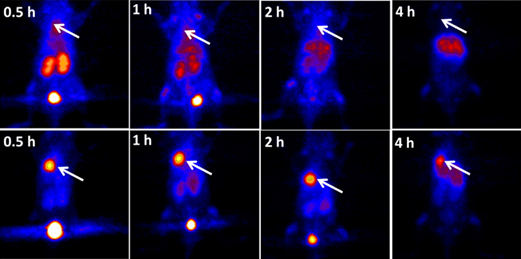
Nude Mouse A549 In situ Lung Cancer Model Imaging