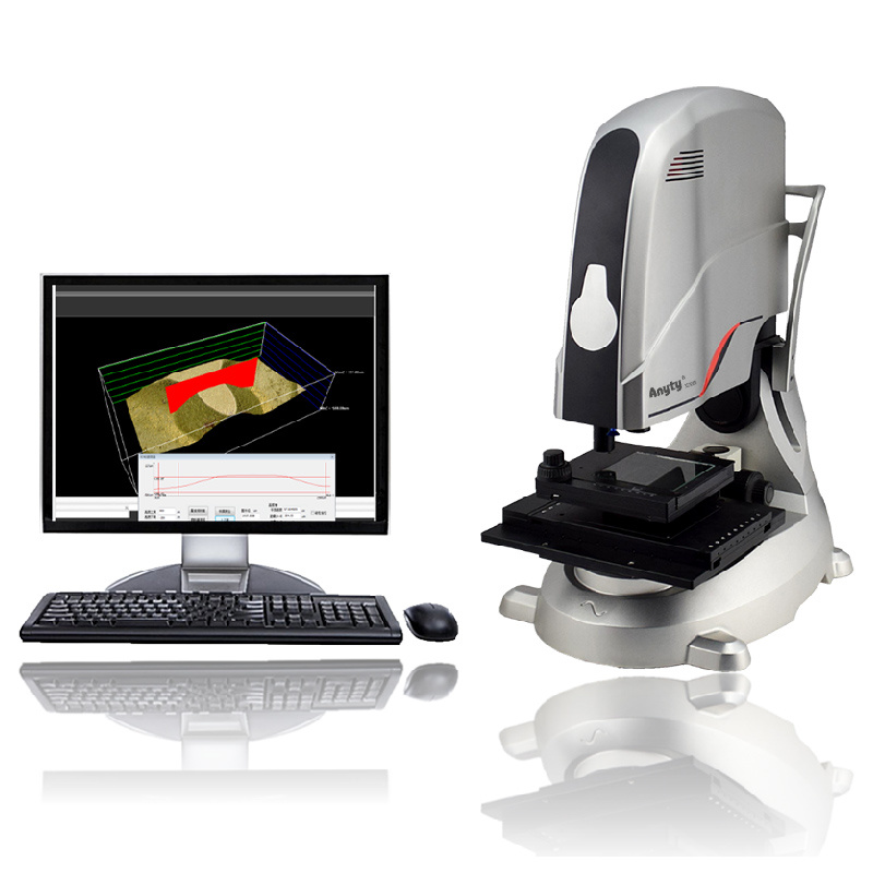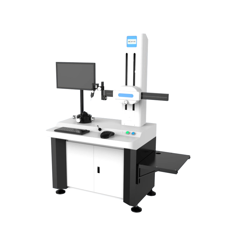


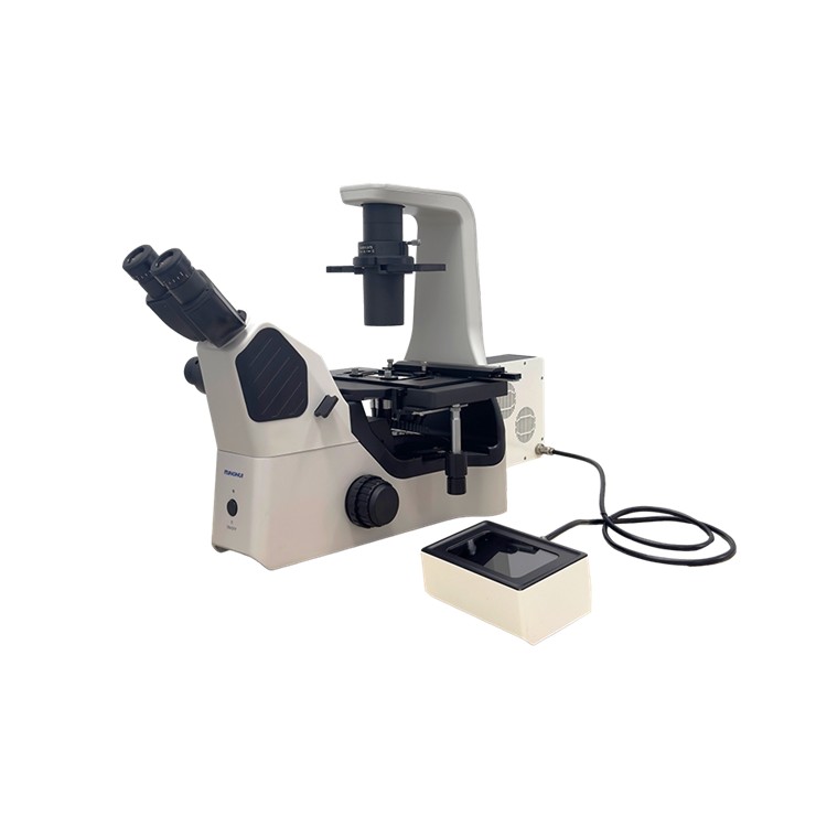
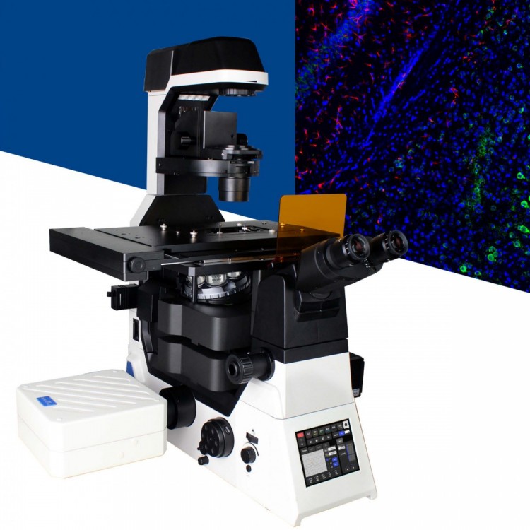
Confocal Laser Scanning Microscope NCF950
The Laser Confocal Microscope NCF950 is a product in the microinstrument series, an essential basic tool designed for laboratory scientific research, offering powerful and stable imaging capabilities and highly integrated电动ization features. The NCF950 is a simple, efficient, and highly integrated domestic confocal microscope. The NCF950 confocal system, with its optical imaging system, straightforward operation, and highly integrated电动ization components, is an indispensable laboratory tool. It is widely applicable, with reliable experimental data and a richly featured analysis and processing software. It plays a role in various fields including cellular and molecular biology, drug screening, neuroscience, immunology, morphology, food hygiene, fermentation, genetics, and pharmacology.
Signal Detection
Efficient scanning heads, detectors, and variable-speed electric micro-holes, combined with Yongxin's powerful optical system, deliver rapid, stable, and high signal-to-noise ratio confocal images.
Multichannel signal detection
Integrated with 4 light sources and detectors (405, 488, 561, 640), combined with 4-channel fluorescence fusion technology, it achieves real-time multi-channel fusion observation and capture.
Electrification hardware
The NCF950 offers a variety of electric motion components, including electric platforms, electric focusing, electric objective rotation disk, electric fluorescence rotation disk, electric condenser lens, and electric dimming. It operates via both button and software controls, and provides command calls for easy user control and development.
Confocal Laser Scanning Fluorescence MicroscopeNCF950Technical Specifications:
| NCF950 Confocal Laser Configuration Sheet
| |
|
Laser pointer
|
Laser wavelengths: 405 nm, 488 nm, 561 nm, 640 nm
|
|
Detector
|
Wavelength: 400-750nm, Detectors: 3 independent fluorescence detection channels; 1 DIC transmission light detection channel
|
|
Scanner Head
|
Pixel Size: 4096 x 4096, Scanning Speed: 2 fps (512 x 512 pixels, bidirectional), 18 fps (512 x 32 pixels, bidirectional), Image Rotation: 360°
|
|
Scanning Mode
|
X-T, Y-T, X-Y, X-Y-Z, X-Y-Z-T
|
|
Pinhole
|
Continuous variable hexagonal electric needle hole; adjustment range: 0-1.5mm
|
|
Co-axial Field of View
|
φ18mm internal square
|
|
Image bit depth
|
12bits
|
|
Accessory Microscope
|
NIB950 All-Electric Inverted Microscope
|
|
Optical System
|
NIS60 Infinite Optical System (F200)
|
|
Eyepiece (Field of View)
|
10x(25), EP17.5mm, adjustable field of view -5 to +5, Φ30mm interface
|
|
Observing Tube
|
Swivel-type trinocular eyepiece tube, 45-degree tilt, interpupillary distance 47-78mm, eyepiece interface Φ30, fixed diopter; 1) Eyepiece/Camera Switch: (100/0, 50/50, 0/100); 2) Visual/Off Visual/Adjustable Focusing Bausch & Lomb Eyepiece
|
|
NIS60 Objective Lens
|
10x achromatic objective lens, NA=0.45, WD=4.0, cover glass=0.17
|
|
20x Achromatic Objective, NA=0.75, WD=1.1, Cover Glass=0.17
| |
|
60× Semi-Apochromatic Objective, NA=1.40, WD=0.14, Cover Glass=0.17, Oil Immersion objective
| |
|
100× Achromatic Objective, NA=1.45, WD=0.13, Cover Glass=0.17, Oil Immersion Lens
| |
|
Objective lens converter
|
Electric 6-way Converter (Expansion Slot), M25×0.75
|
|
Converging lens
|
6-hole electrical control: NA0.55, WD26; phase contrast (optional: 10/20, 40, 60)
|
|
DIC (10X, 20X/40X) Optional. Empty hole
| |
|
Lighting System
|
Transmitted Collar Lighting, 10W LED Lighting
|
|
Projector Lighting: Wide Field Fiber Optic Lighting 6-hole electric fluorescent turntable (B, G, U standard); electric fluorescent light gate;
| |
|
Mid-Ratio Switch
|
Manual 1X, 1.5X, Coaxial Switching
|
|
Airframe port
|
Spectral ratio:
|
|
Left: Visual = 100:0; Right: Visual = 100:0;
| |
|
Platform
|
Electric Control: Stroke range 130mm x 100mm (tabletop 325mm x 144mm); Speed: 25mm/s; Resolution: 0.1μm - Repeatability: 3μm. Mechanically adjustable sample clamps
|
|
Focusing System
|
Coaxial coarse and fine lifting mechanism, stroke: 7mm up, 2mm down; coarse adjustment 2mm/rev, fine adjustment 0.002mm/rev; manual and electric control available, with electric control, minimum step of 0.01um.
|
|
DIC plug-in board
|
10X, 20X, 40X plates; fits in converter slots; optional
|
|
Control
|
Joysticks, control panels, USB cables
|
|
Software
|
Software: NOMIS Advanced C Image Display/Processing/Analysis 2D/3D/4D image analysis, temporal variation analysis, 3D image acquisition and orthogonal display, image stitching, multi-channel color confocal images
|
Confocal Fluorescence Microscope, Guangzhou Confocal Microscope, Laser Confocal Microscope, Confocal Laser Microscope, Laser Confocal Fluorescence Microscope, Confocal Scanning Microscope