


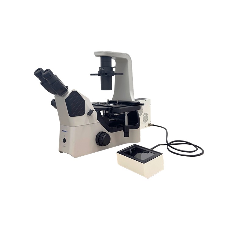
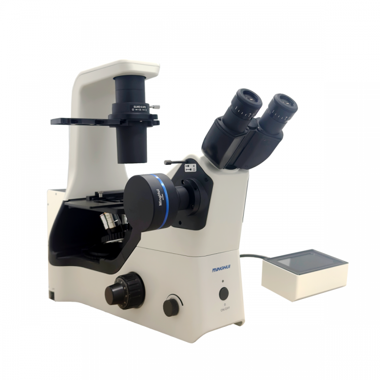


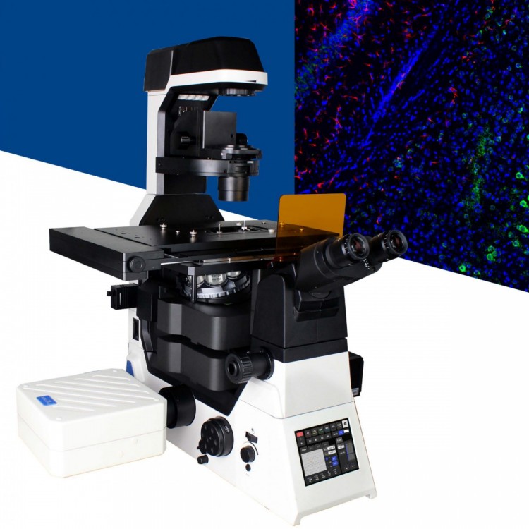
Product Description
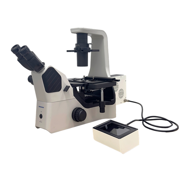
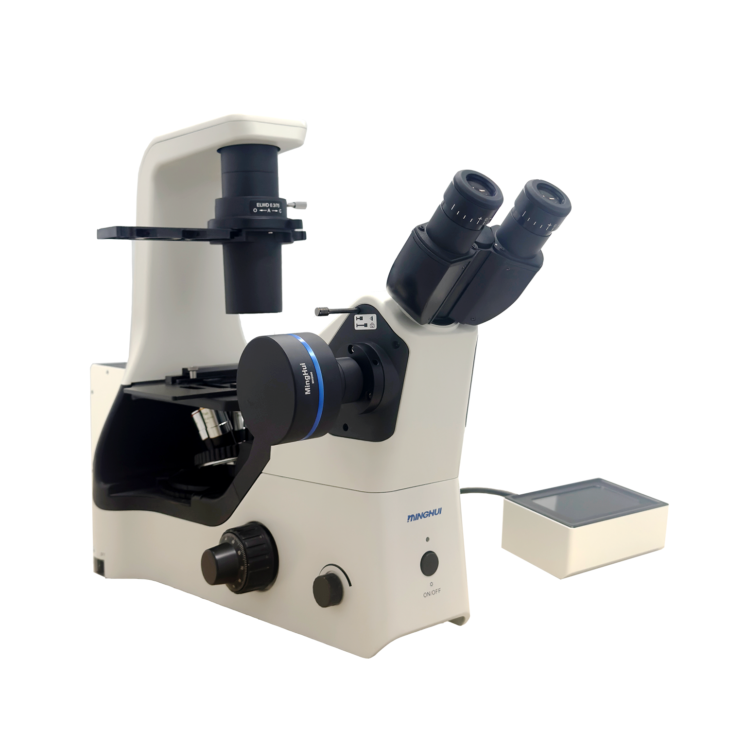
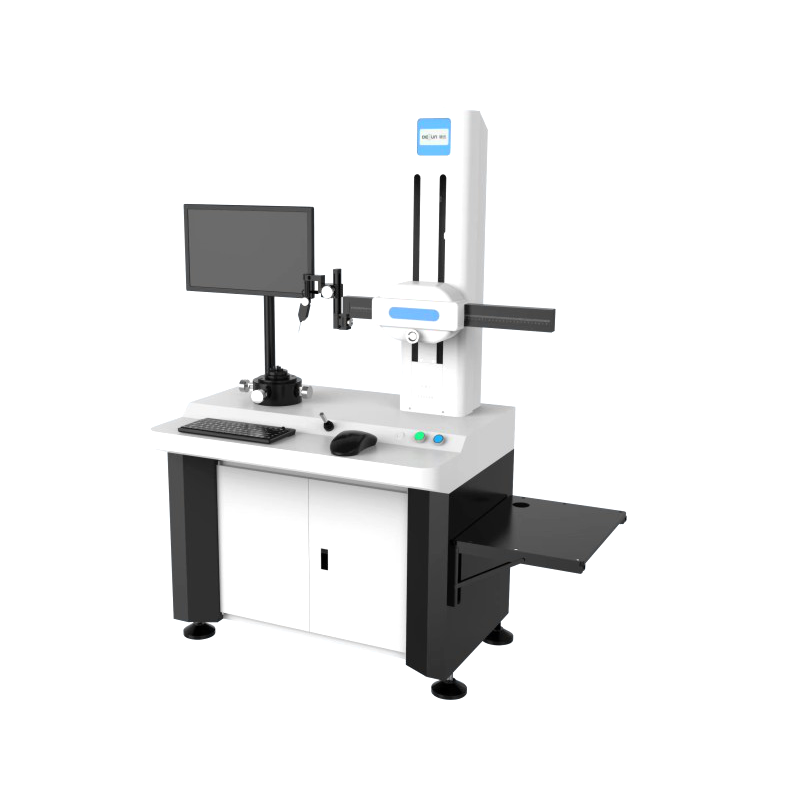
MHIF3000Research-grade inverted fluorescence microscopes utilize an infinite conjugate flat field semi-apochromatic optical system, equipped with a swing-out trinocular observation head and a flat field semi-apochromatic objective lens group, supporting bright field, phase contrast, and fluorescence multi-mode observations. Its flat specimen stage is compatible with dishes, multi-well plates, and other containers, paired with a long working distance condenser and a universal phase contrast slide, meeting the needs for dynamic observation of living cells. The fluorescence system employs6Workstation manual turntable design, standard configuration385nm/460nm/520nm/625nmFour Channel 75WHigh-powerLEDFluorescent light source, supporting multi-color synchronous imaging, performance comparable to imported equipment. The overall modular structure balances high-resolution imaging and experimental expandability, suitable for advanced scientific research scenarios such as cell biology and drug screening.
Product Specifications
Optical System | Infinite Field Semi-Apochromatic Optical System | ||
Observation System | Swivel three-way, 0:100% two-speed split light | ||
Observation Method | Bright Field, Contrast, Fluorescence | ||
Eyepiece | 10X adjustable eyepiece, line field of view Ø22 | ||
Objective Lens | Flat Field Semi-Apochromatic Differential Contrast Fluorescent Objective | ||
4XPH 4X Objective Lens 4XPH NA 0.13 W.D. 10.43mm | |||
10X PH Objective, 10X Magnification, NA 0.25, W.D. 7.3mm | |||
10XFL 10X Semi-Plan Fluorescence Objective 10X Fluor NA 0.30 W.D. 8.31mm | |||
20XFLPH 20X Semi-Plan Achromatic Fluorescence Objective 20X Fluor PH NA 0.45 W.D. 7.11mm | |||
40XFLPH 40X Semi-Plan Achromatic Fluorescence Objective 40X Fluor NA 0.65 W.D. 1.6mm | |||
Light Source | 3W S-LED lighting with adjustable brightness | ||
Converter | 5-Pin Objective Converter | ||
Loading Platform | Flat loading platform: 170(X) × 250(Y) mm, equipped with replaceable droplet loading plate (φ110), optional extended tray | ||
Mechanical Moving Scale, Travel: 129 (X) x 83 (Y), compatible with five micro experimental boards, porous plate clamps, and specimen stage clamps, equipped with a universal tray. | |||
Suitable for Terasaki plates, slides, Φ35-65 petri dishes, and Φ35mm petri dishes, among various holders. | |||
Focusing Method | Coarse and fine adjustment of coaxial focusing; right-hand side features coarse adjustment tension control; fine adjustment 0.002mm/step, 0.2mm/revolution | ||
Rough movement 37.7mm/circle, objective turret vertical travel: top 7mm, bottom 1.5mm, maximum 18.5mm with limit disengaged | |||
Concentrating lens | Long-focus condenser lens, numerical aperture 0.3, working distance 75mm; removing the condenser lens allows for a working distance of 187mm. | ||
Matching Device | The focusing lens is equipped with 4X, 10X, 20X, and 40X universal phase contrast slides. | ||
Fluorescent Device | Equipped with a multi-functional rotary disk structure, 6-position manual rotary disk; up to six fluorescence excitation modules can be installed as needed. | ||
Standard with three sets of fluorescence filter blocks | FL-UA | UV Bandpass UA: 365/30nm DM: 405nm EM: 460/50nm | |
FL-BA | Blue Bandpass BA EX: 470/30nm DM: 505nm EM: 535/35nm | ||
FL-G | Green Long Pass G EX: 530/30nm DM: 565nm EM: 575nm LP | ||
High-power long-life fluorescent LED light source | Standard configuration with 385nm/460nm/520nm/625nm four-channel output, 75W high-power output, available for secondary development | ||
Imaging System | USB2.0 | MHD500 | |
USB3.0 | MHC600、MHD600、MHD800、MHD1200、MHD2000、MHS500、MHS900、MHS2100 | ||
Inverted Fluorescence Microscope MHIF3000 Application Fields
Section 1: Biomedical Research
Live Cell Observation
•Supports real-time dynamic observation of cell culture, explant tissues, and plankton, with long working distance objectives allowing direct microscopic analysis of samples in petri dishes.
• Multi-channel fluorescence synchronous imaging function (e.g., 385nm/460nm/520nm/625nm, four channels) is suitable for immunofluorescence labeling and organelle localization studies.
2. Pathology and Drug Development
• High-resolution imaging in cancer therapy nanomedicine research, supporting drug mechanism verification and cell transfection process monitoring
• Smart algorithms ensure clear image capture in low-light conditions, meeting the requirements for染色细胞 observation and pathological diagnosis.
2. Industrial and Materials Science
• Modular design compatible with bright-field/fluorescence/differential interference contrast multimodal imaging, expandable for surface defect detection on coatings, thin films, etc.
• Achieve visual analysis of heterogeneous material interfaces through pseudo-color synthesis technology
Section 3: Training and Quality Control
• Used for teaching and demonstration in university laboratory cell biology, a single unit can efficiently complete large-scale sample testing
Microstructural analysis plays a role in food inspection and water quality assessment, catering to the needs of research institutions and inspection and quarantine agencies.
The equipment ensures experiment reproducibility through OLED screen parameter display and brightness memory function, and its performance is on par with imported high-end models.