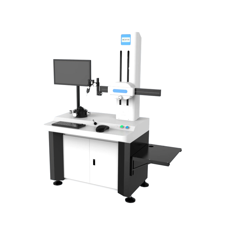


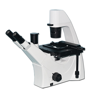


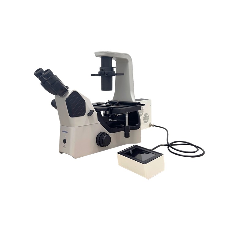
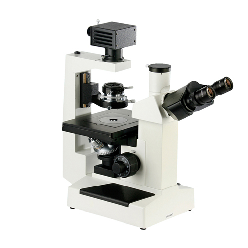
MHIL150 Inverted Optical Microscope, designed specifically for cell tissue culture observation, featuring high contrast and resolution to highlight cell contours and internal structures, meeting the needs of laboratory daily operations and fundamental research. Paired with a high-sensitivity, high-resolution microscope camera, it allows for digital imaging observation when connected to the microscope. Offers 5 to 20 million pixel camera options to accommodate various types of microscopes and their imaging software. Connectable to a computer for image capture, storage, processing, analysis, and sharing. Provides SDK for secondary development and lifetime free software upgrades.

Inverted Optical Microscope MHIL150
The purpose of an inverted microscope:
1. Observe Living Cells: Inverted biological microscopes are suitable for observing living cell cultures. As the inverted objective lens allows direct observation of cells at the bottom of the cell culture dish, it is possible to witness the growth and division processes of the cells.
2. Observe Multilayer Tissue: An inverted biological microscope allows for the observation of the structure of multilayer tissue, as the inverted objective lens can directly view the tissue surface without the need for tissue sectioning.
3. Observation of Aquatic Organisms: Inverted biological microscopes are suitable for observing aquatic organisms, as the inverted objective lens allows direct observation of organisms in water without the need to remove them and place them on a microscope slide.
4. Observe Large-Scale Samples: As the lens of the inverted biological microscope is positioned above the object, it allows for the observation of a larger area and thicker samples compared to conventional microscopes.
5. Observe Sample Changes: Inverted biological microscopes are suitable for extended observation, enabling the viewing of cellular growth and changes, which aids in studying the physiological and biochemical processes of cells.
| Inverted Biological Microscope MHIL150 Configuration Sheet
| |||||
|
Key Parameters
|
Total Magnification
|
100X~400X (Standard Configuration)
| |||
|
Mechanical cylinder length
|
∝
| ||||
|
Objective conjugate distance
|
∝
| ||||
|
Objective Lens
|
Wide-field eyepiece for horizontal field of view
|
WF 10X
|
Field of View: Φ22mm
|
Eyepiece Interface Ф30mm
|
Collimation Distance 10mm
|
|
Chinese telescopes
| |||||
|
Eyepiece Tube
|
45-degree tilt, binocular interpupillary distance adjustment range: 48~75mm, eye point height from the work surface is 400mm.
| ||||
|
Clear Field Objective
|
Magnification Ratio
|
Numerical Aperture
|
Working Distance (mm)
|
Cover glass thickness (mm)
|
Focal Length
|
|
4X
|
0.1
|
21
|
0.
|
50
| |
|
Matching Objective Lens
|
10X
|
0.25
|
4.3
|
0.
|
20
|
|
20X
|
0.4
|
8
|
0.
|
10
| |
|
40X
|
0.6
|
3.5
|
0.
|
5
| |
|
Converter
|
Four-hole converter
| ||||
|
Focusing Mechanism
|
Coarse-fine coaxial, fine adjustment value: 2μ, coarse adjustment tension adjustable, with locking and limit devices, effective focusing travel 11mm
| ||||
|
Converging lens
|
Long working distance objective lens, working distance 70mm, with a plate-type phase contrast device
| ||||
|
Loading Platform
|
Motion Range (Horizontal X Vertical): 112mm x 79mm, the motion scale is detachable
| ||||
|
Petri dish tray
|
86mm (width) x 129.5mm (length), compatible with round culture dishes Φ87.5mm
| ||||
|
34mm (width) x 77.5mm (length), compatible with circular culture dishes with a diameter of Φ68.5mm
| |||||
|
57mm (width) x 82mm (length)
| |||||
|
Complementary System
|
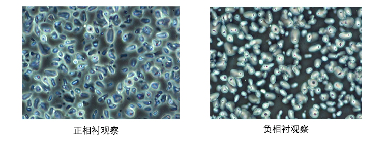
Phase Contrast
|
Lever-actuated compensating condenser with adjustable center contrast ring
| |||
|
Lighting System
|
9W LED, adjustable brightness
| ||||
|
Color Filter
|
Matte glass, blue filter sheet
| ||||
|
Adjustment Tools
|
Hexagon socket wrench (M4, M5)
| ||||