


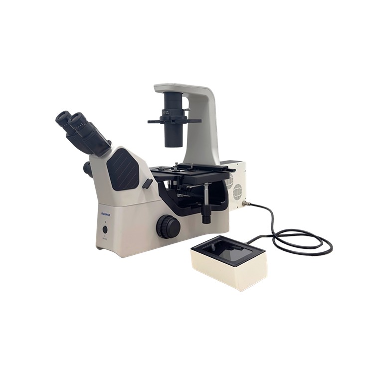
Incorporating pathological studies into higher-level cellular research areas distinguishes inverted microscopes from traditional upright microscopes, which are primarily used for pathological section observation. Inverted microscopes are more commonly applied to microscopic observations at the cellular level. Medical research today no longer confines itself to macroscopic physiological studies but has entered the cellular domain, understanding the human body from genetic and molecular perspectives, and conducting medical treatments and resolving medical challenges.
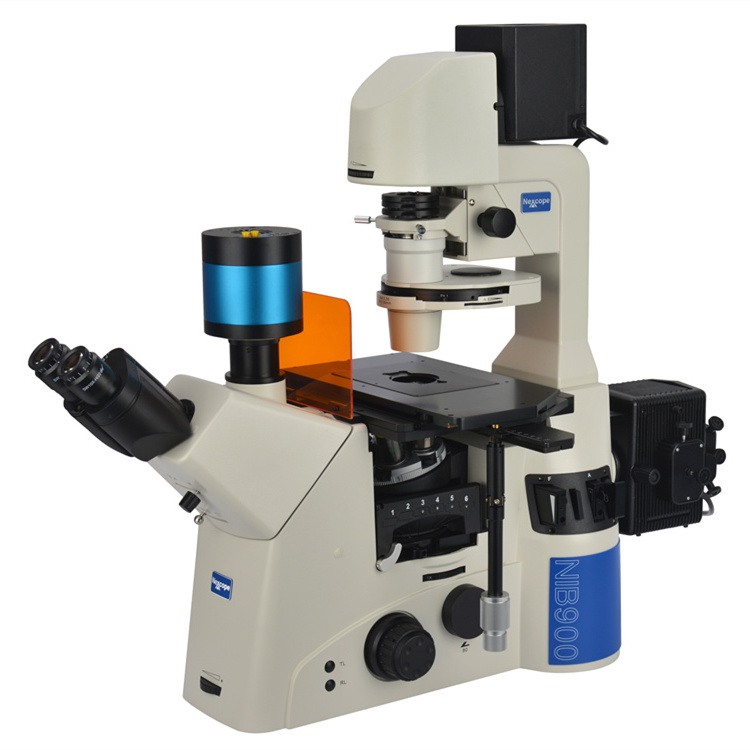
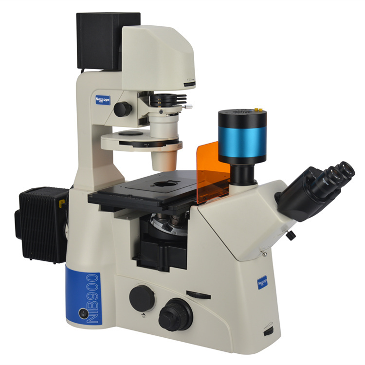
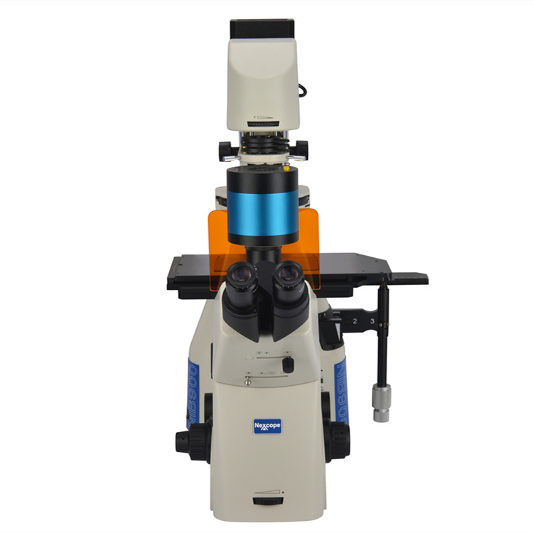
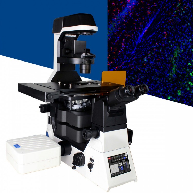
The NIB900, a research-grade inverted microscope designed to meet the advanced needs of life science research, caters to your diverse requirements. It is a versatile microscope capable of bright-field, dark-field, phase-contrast, polarized light, DIC, and fluorescence observations. It can even be upgraded to meet the needs of confocal and super-resolution microscopy for life science research.
The NIB900 is also compatible with an external microinjection system, a technology that has become a very common operational method in the field of medical and biological research. It allows for nuclear transplantation, microinjection, somatic fusion techniques, embryo transfer, and micro-cutting, as well as operations in emerging research technologies like transgenesis. This microscope is an indispensable part of a pathology laboratory, serving as the right-hand man and good companion to pathologists.
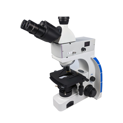
Scientific Inverted Fluorescence Microscope NIB900-FL - Brightfield, Darkfield, Phase Contrast, Polarized Light, DIC, Fluorescence
1. Main Functions: Suitable for various observation methods including bright field, dark field, polarized light, phase contrast, and DIC.
2. Optical System: Infinite Field (Tubular Microscope 200) Flat Field Semi-Aplanatic Optical System
3. Observation System: Hinge-type Trinocular (with Bausch & Lomb eyepiece), B:T = 50%:50%, 0:****, ****:0; Pupillary Distance 55-75mm
4. Eyepiece: 10X adjustable diopter, line field of view Ø22 WF10X/Ø22
5. Objective Lens: Plan Apochromatic Contrast Objectives with semi-reduced chromatic aberration, delivering high signal-to-noise ratio, high resolution, and high contrast imaging under various illumination modes.
10X NA=0.30 WD=8.1 Cover Glass Thickness: 1.2
20X NA=0.45 WD=7.5-8.8 Cover Glass Thickness: 0-2 mm with Correction Ring
40X NA=0.60, WD=3-4.4, Cover Glass Thickness: 0-2, with Correction Ring
6. Converter: 6-hole objective converter with DIC slot (suitable for both transmitted and reflected light).
7. Load Platform: Three-tier mechanical moving platform with a movement range of 130x85mm, featuring a flexible handle. Different-sized small workbenches can be installed on the top level according to requirements.
8. Focusing Method: Coaxial coarse and fine adjustment lifting mechanism, travel range of 9mm (upward 2mm, downward 7mm), fine adjustment range of 0.1mm per revolution, fine adjustment reading of 1um.
9. Focusing Mirror: Long working distance turntable design, 6-position turntable (1 bright field, 3 phase contrast, 2 DIC), NA 0.55, WD=26mm.
10. System Interpolation Ratio: 1X, 1.5X.
11. Side End Outlets: Rotating disc structure (manual). Left / Visual = **** / 0%; Right / Visual = 20% / 80%; Both Sides / Visual = 0 / ****:
12. Lighting System: Kohler Lighting, 12V 100W Halogen Light Room
13. Designed for easy handling with a convenient handle placement.
Structure Type: Transmission Lighting/Epifluorescence Microscope