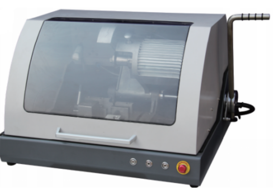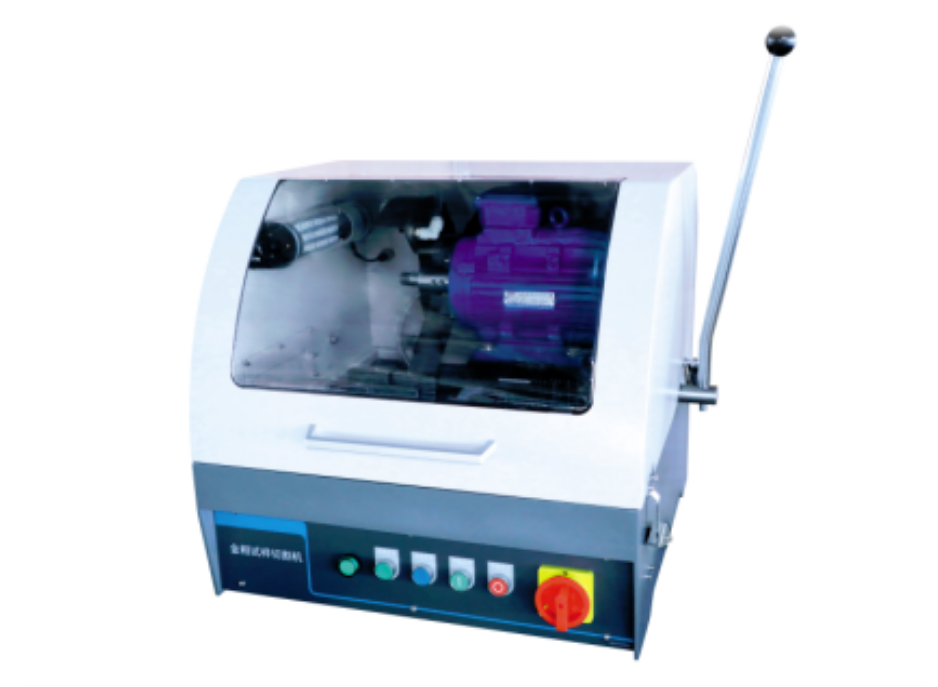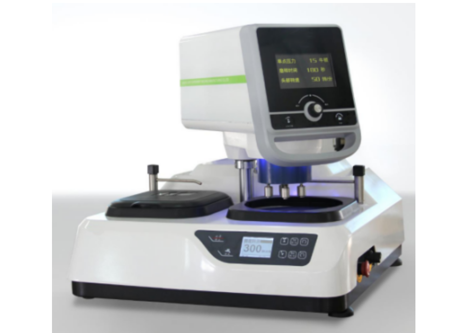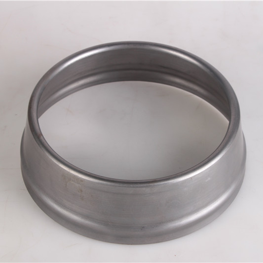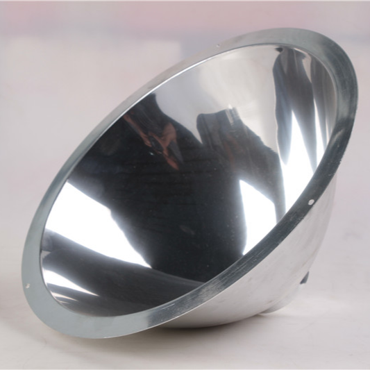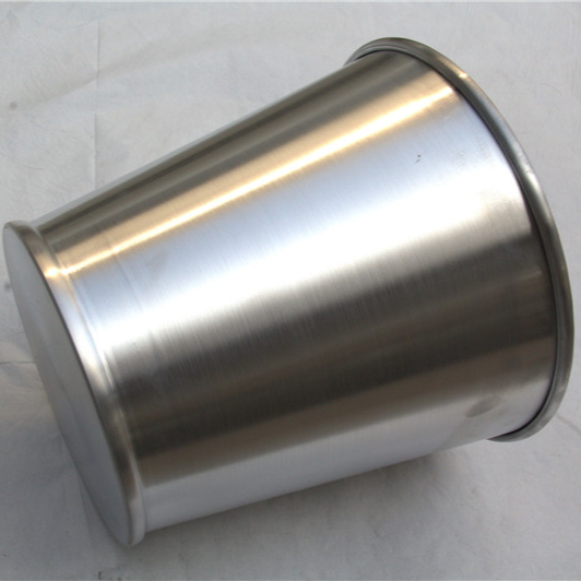


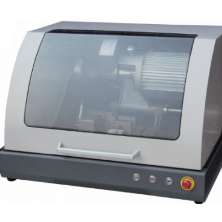


OneStructural Description
1) Appearance
2) Size(mm):

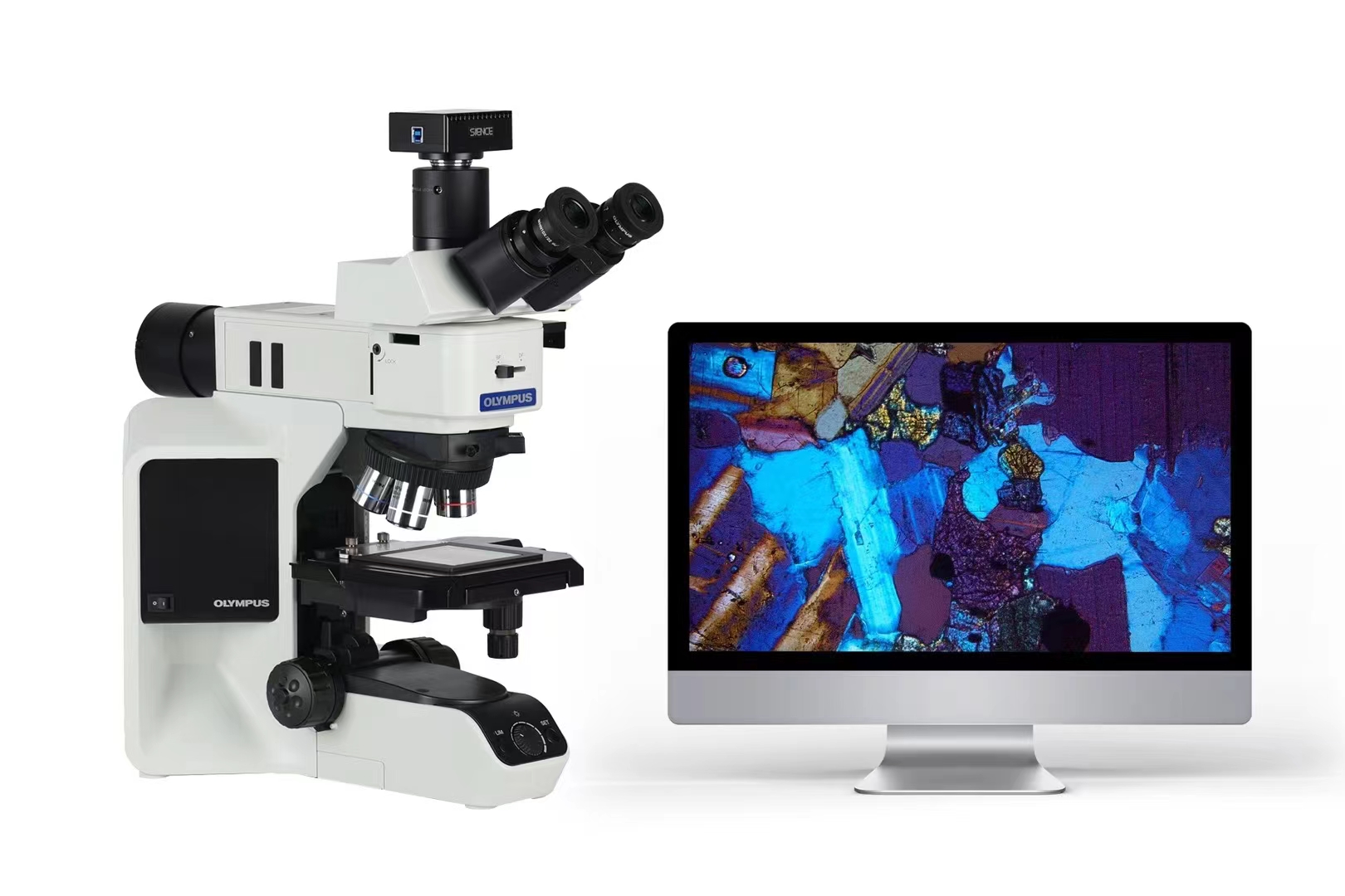
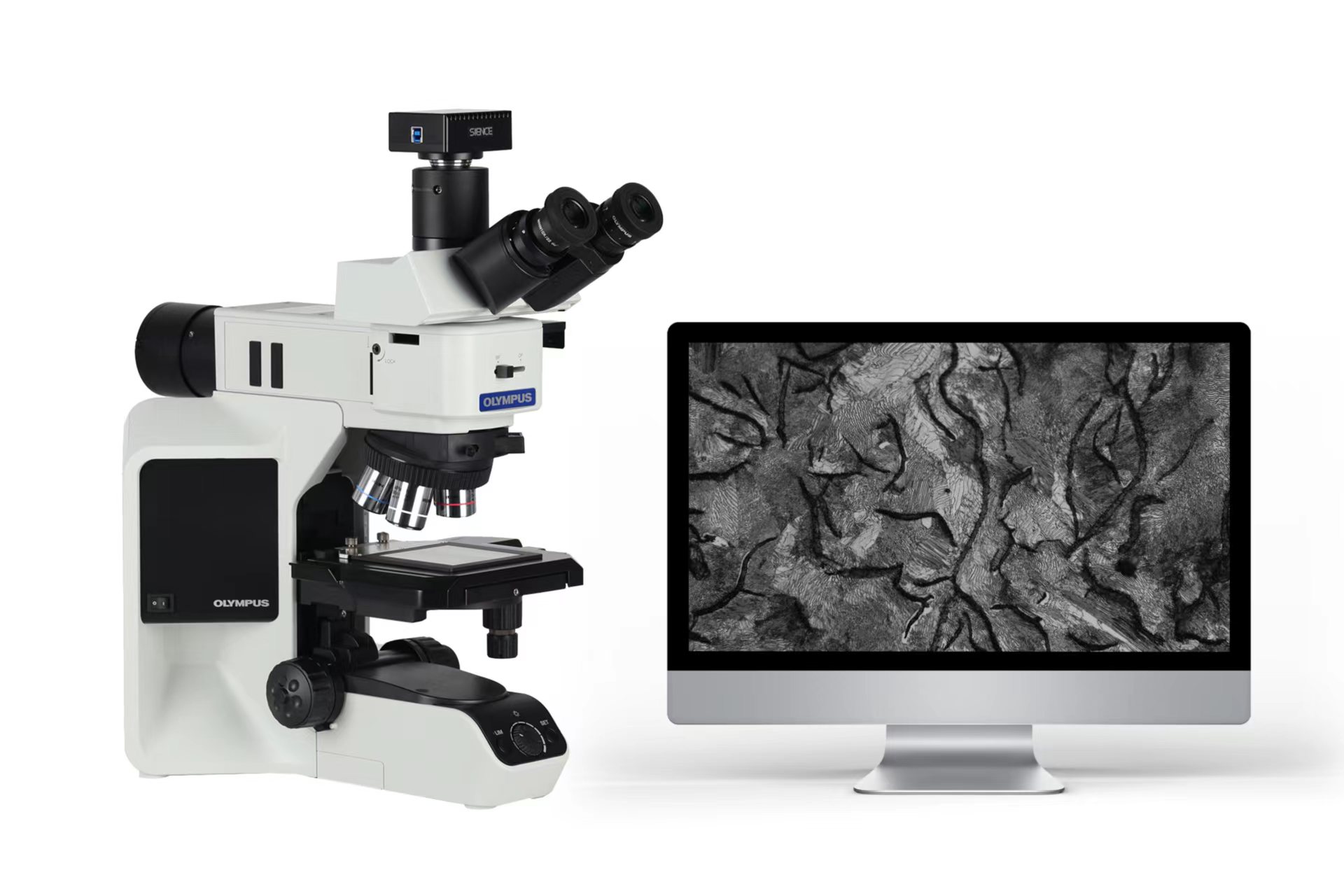
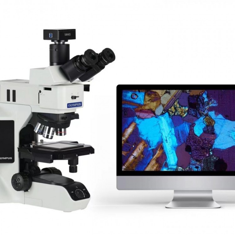
TwoSystem Composition
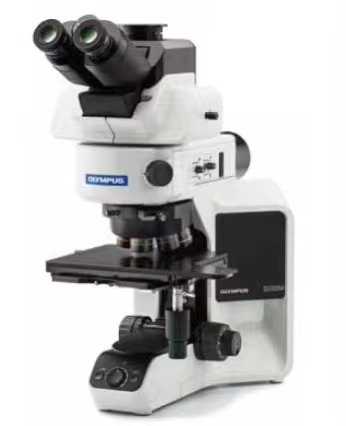
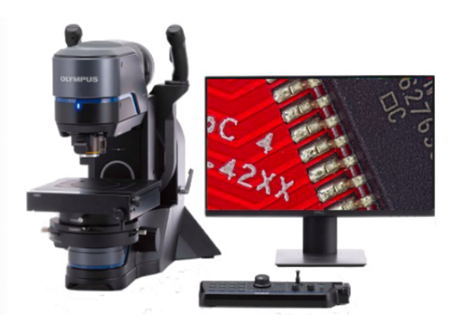
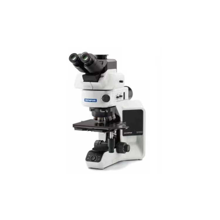
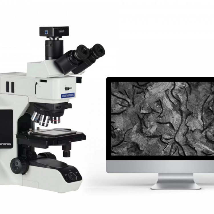
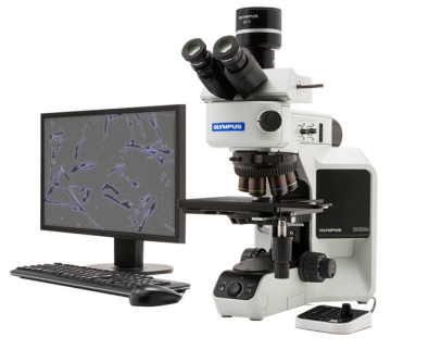
1) Vertical Metallographic MicroscopeBX53M;
2) Microscope Digital Camera3.0Interface;
3) Metallographic Analysis Software;
4) Computers (Dell desktops, domestically supplied)
ThreeTechnical Advantages
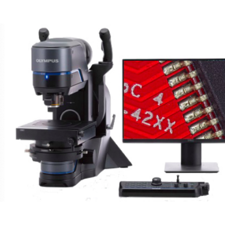
BX53MIs from Olympus Corporation of Japan2016Announced this year, the new intelligent upright metallographic microscope. An integrated solution that combines a microscope, camera, and software, guiding users through the entire workflow. Directed darkfield,MIXLighting and other functions, enablingBX53MNow an advanced upright metallographic microscope in the industry. With the powerful OlympusStreamCombining software, we can offer users dozens of solutions.
1) High-quality optical systems, providing excellent imaging quality. Objective lenses with wavefront aberration control technology offer improved resolution compared to conventional lenses.
2) Smart light intensity management. Allows for independent setting of appropriate lighting intensity based on different magnification levels and observation modes.
3) Brand-new high-brightness white light designLEDLight source, super long lifespan, maintains a consistent color temperature.
4) Can preset working modes for different samples without needing complex adjustment settings each time. One-click restore to working mode ensures efficiency is not compromised even with multiple users.
5) Olympus Directional DarkfieldDDFFunction: View samples from multiple angles to ensure ideal observation effects.
6) OlympusMIXLumination, combining various observation modes to create rich new observation patterns.
7) A wide variety of objective lenses are available, in addition to those with standard magnification ratios, we also have...1.25XAnd2.5XUltra-low magnification objective150XHigh magnification objective lens40XObjective lenses are available for selection.
8) A micrometer-adjustable aperture stop and field stop with scale indicators, allowing even beginners to easily set the aperture size according to different observation conditions.
9) Combined with software, achieve automated management.
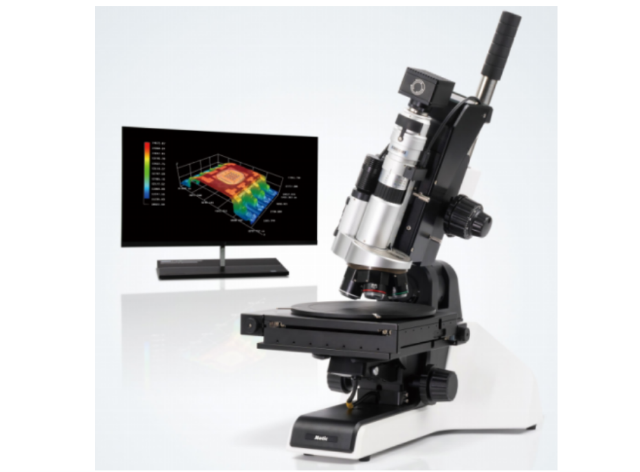
10) Integrated diffractive interferometer design; the background interference color changes continuously from gray to magenta sensitive color through adjusting the control knob, allowing the interference color to be set for contrast, presenting a bas-relief-like appearance.3DImage.
11) Innovative focusing mechanism design significantly enhances service life.
12) All optical components are treated with a patented anti-mold technology, ensuring they do not mildew over long periods of use.
V. Key Parameters
1) Microscope Type:
Vertical metallographic microscope
2) Objective Lens:MPLN5X,MPLN10X,MPLN20X,MPLN50X,MPLN100X;
3) TotalMagnification Ratio:
50×、100×、200×、500×、1000×;
4) Light Source Type:
High-lumen white lightLEDLight source, super long lifespan, consistent color temperature.
Reflective light/Transmitted light
5) Eye Field of View:
22mmWide field of view; with diopter compensation, pupillary distance adjustment55~75mm);
Reticle Eyepiece Micrometer with Crosshair10mm100Equally divided (or equally spaced)
6) Observation Method:
Field of view, dark field, polarized light, differential interference, directional dark fieldMIX;
7) Smart Light Intensity ManagementLIM)
The body features an integrated light intensity control button, allowing for the adjustment of appropriate brightness levels for different observation modes or magnification settings.
8) Focusing Mechanism
Focusing Travel:25mm
Micro-tuning scale:1um;
Focusing limit protection mechanism, coarse adjustment knob tension adjustment mechanism.
9) Sample Height
65mm(Standard)/105mm(with height adapter)
10) Optical Path Interface
Simultaneously external2Image recording equipment
Three-section spectrometer, ratio100:0、20:80、0:100;
11) Objective lens turret:
5Holes, with positional coding, electrically adjustable.
Open Field/Dark field general use.
12) Focusing Mechanism
Precise Coarse/Fine-tuning focusing mechanism, with anti-slip mechanism, upper limit safety protection mechanism, and tension adjustment mechanism.
The body features focusing scale indicators to guide the focusing direction.
13) Loading Platform
High precisionXYSliding Mechanical Load Platform
Coaxial Left/Right-hand operated mechanical loading platform: Stroke76×52mmTorque adjustment
Large Left/Right-hand operated mechanical loading platform: stroke100×105mm, withYAxle Locking Device
Large coaxial right-hand operation mechanical load platform: stroke150×100mmWith torque adjustmentYAxle Locking Device
14) OptionalObjective Type: Plane Field Semi-Apochromatic Objective; made of Fluorite material, stress-relieved and coated to prevent mildew.
Model | Magnification Ratio | Working Distance | Numerical Aperture | Resolution |
MPLN5XBD | 5Multiple | 12.0mm | 0.15 | 2.24um |
MPLN10xBD | 10Multiple | 6.5mm | 0.30 | 1.12um |
MPLN20xBD | 20Multiple | 3.0mm | 0.46 | 0.73um |
MPLN50xBD | 50X2 | 1.0mm | 0.80 | 0.42um |
MPLN100xBD | 100Multiple | 1.0mm | 0.90 | 0.37um |
15) Imaging Record Equipment:UM 1000Microscope-specific digital camera
l High-definition, high-sensitivityUSB3.0InterfaceEffective Pixels1000Ten million, non-interpolation operation.
l True color reproduction, completely eliminating the drawback of unsatisfactory color reproduction in common digital cameras.
l High-sensitivity photo sensors capable of capturing low-light dark field images.
l Utilizing high-speedIEEE1394Transmission method, continuous image without latency, capable of real-time acquisition, synchronized capture of dynamic images, atLCDThe screen display is visible, and can also be observed through the eyepiece, with easy focusing and browsing.
l Photos can be saved in various file formats.TIF, BMP, JPG, PSD, GIF…), compatible with various general image processing programs.
l The photoelectric chip features an automatic protection system, stable operation, and an exceptionally long service life.
1) Metallographic AnalysisSoftware, all hardware parameters, select the corresponding hardware to obtain the corresponding scale, no need for cumbersome calibration operations.
2) Pre-set microscope parameters (brightness, aperture, objective magnification) with one-click recovery (hardware support required).
3) Multilingual operational interface
4) Interactive measurement feature, offering a variety of measurement tools.
5) Data can be output toExcelTable, or save overlayed on the image together.
6) Customizable report formats can be generated, with reports ready for direct use in Microsoft applications.OFFICESoftware Editor
7) Robust image processing capabilities, one-click operations for shadow correction, contrast enhancement, etc. ;
8) Real-time Depth of Field Expansion FeatureLive EFI),Easily capture images of samples with slopes or uneven surfaces;
9) Panoramic Photo Puzzle(Live MIA)Automatically performs image stitching while moving the sample, expanding the field of view of the microscope.
10) The phase analysis function allows for quick and accurate determination of multi-phase area content.
11) Available with a variety of material testing solutions, including grain size grading, cast iron analysis, particle analysis, porosity measurement, and layer thickness measurement, etc.