

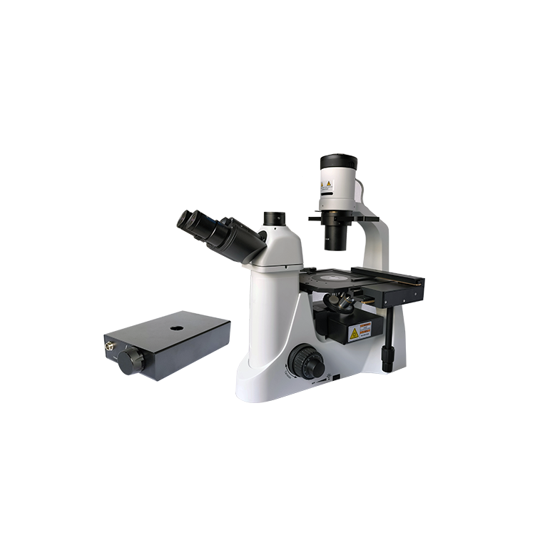
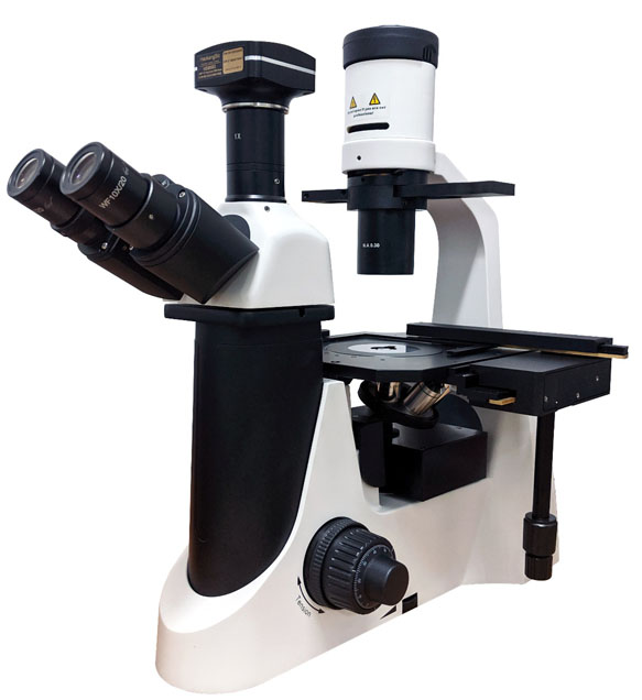
Inverted Fluorescence Microscope MHIF2000
Inverted microscopes are widely used in medical testing, pathological diagnosis, immunofluorescence, and cell observation fields. They are utilized for observing and researching biological sections, cells, bacteria, and living tissue cultures, as well as fluid precipitates. Additionally, they can observe other transparent or translucent objects, powders, and fine particles. They are applied in scientific research institutions, universities, healthcare, inspection and quarantine, agriculture, animal husbandry, and dairy industries, where they play a significant role in food inspection, water quality analysis, crystal structure analysis, and chemical reaction precipitate analysis. This product can be customized with a fluorescence attachment to enable fluorescence observation, allowing for easy switching between brightfield and fluorescence modes via a pole or turntable. It also connects to cameras via an interface adapter for microscopic imaging purposes. Moreover, it boasts excellent performance at a reasonable price.
Product Specifications
Optical System | Infinite color difference independent calibration optical system | Quantity | |
Configuration Type | Fluorescent | / | |
Eyepiece | High eye point flat field eyepiece 10X/Φ20mm | 2 | |
Focusing Mechanism | Adjust the coarse/micro focus knob to control the vertical movement of the objective disc, travel: 14mm, equipped with a limit switch and locking device. | 1 | |
Low position coarse and fine adjustment coaxial focusing handwheel, each turn focus knob stroke: 20mm (coarse adjustment), 1μm (fine adjustment) | |||
Objective lens turret | Five-hole objective rotator | 1 | |
Loading Platform | Flat loading platform: 200mm (L) x 266mm (W), equipped with replaceable water drop loading plate (φ110) | 1 | |
Water Droplet Carrier Sheets + Compression Set | |||
Matching device | Matching Ring Plate Sliding Assembly | 1 | |
Converging lens | Long working distance condenser lens, rotary phase contrast device, phase contrast observation adaptation | 1 | |
Numerical Aperture: 0.30; Working Distance: 72mm | |||
Applicable Objective: 2x to 60x, with an aperture stop | |||
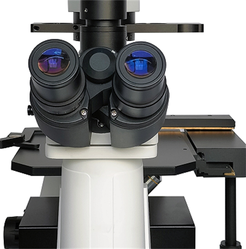
Observing Tube | Three-axis observation tube, 30° tilt, 360° rotation, pupillary distance 48-75mm, ±5 diopter adjustment | 1 | |
Optic Path: Eyepiece/Imaging Port = 0:100/100:0, switched via the pull rod | |||
Objective lens | Infinite Far Field Flat Field Achromatic | 1 | |
·10X NA 0.25 W.D. 9.5mm | |||
·20X NA 0.40 W.D. 7.97mm | |||
·20X PH NA 0.40 W.D. 7.97mm | |||
·40X NA 0.60 W.D. 3.77mm | |||
Fluorescent illuminator | LED light source, brightness adjustable, with digital intensity display, 12V/2A power adapter | 1 | |
Standard with three sets of excitation blocks; others are customizable. | UV: EX360/50nm; DM: 400nm; EM: 410nm LP | ||
Blue (B): EX475/35nm; DM: 500nm; EM: 530/50nm | |||
Green (G): EX530/40nm; DM: 560nm; EM: 575nm LP | |||
Imaging Port | C-type interface; TV1XC-U | 1 | |
Top light source | 5W LED source, continuously adjustable | 1 | |
Attachment | Dust cover | 1 | |
Rated Voltage/Current | AC 100-240V 50/60Hz 0.4A | 1 | |
Application Fields
