

The NCF1000 Laser Confocal Microscope features an industry-leading 25mm field of view, enabling unobstructed coverage of large sample areas for seamless wide-field imaging. With a scanning size of 8192 x 8192 pixels, it achieves vivid and detailed imaging even with low magnification. Its exceptional imaging capability allows for high resolution on a single image, collecting more and faster data per image while satisfying wide-field observation.
Wider field of view, higher resolution, and faster speed mean you can achieve superior image quality, clearer contrast, and more realistic colors. Lead you to delve into the nanoscale world within cells, interpret cellular reaction processes more intuitively and accurately, and help expand the boundaries of your scientific research, continuously exploring the unknowns in life sciences.
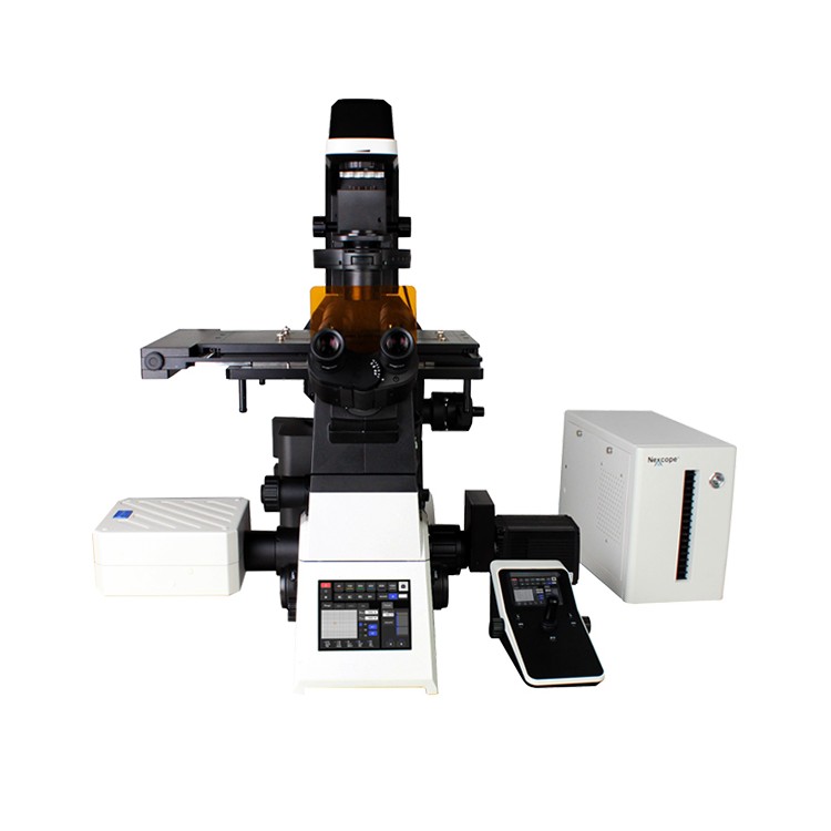
NIB1000 offers a robust and flexible imaging solution, establishing a solid and highly scalable foundation for the NCF1000 series of confocal systems. The 25mm field of view design provides ideal observation conditions for large sample, high-throughput experimental research. Combining bright field, fluorescence, differential interference contrast, and phase contrast microscopy, users can freely choose between single or double-layer optical path configurations based on specific experimental content to achieve optimal imaging results. The Adaptive Focus Shift System (AFS) ensures precise focal plane location during continuous observation, resulting in stable, continuous, and clear recordings of cellular dynamic behavior.

Supports ultra-resolution module
Multimodal
> Stage, Contrast, Fluorescence, DIC
Laser Confocal
>2D-SIM/3D-SIM/TIRF-SIM
2. Ultra-wide view, ultra-high resolution, ultra-fast speed
Viewing Range: 133 μm x 133 μm (100%)
Horizontal resolution (2D-SIM) doubled.zuiUp to 85nm
Axial hyper-resolution (3D-SIM) improved to 270nm
The A1 module can boost resolution up to 60nm.
Laser Confocal Microscope NCF1000 Product Specifications
| Category | NCF1000 | NCF1000 - Lite |
| Laser | Laser wavelengths: 405nm, 488nm, 561nm, 640nm | |
| Standard Detector | Wavelength: 400 - 750nm, 4-channel PMT detection | Wavelength: 400 - 750nm, 1-channel PMT detection |
| Spectral detector | Wavelength: 400 - 750nm, 4-channel PMT detectionzuiMulti-compatible with 4-channel GaASP PMT detection, spectral range 400-750nm, adjustable accuracy 1nm. | - |
| Transmission Detector | Single-Channel PMT Detector - | |
| Scanner Head | zuiLarge pixel size: 8192 x 8192 | |
| Scan Mode | Support for combined usage of scanning functions for X, Y, Z, λ, T | |
| Pinhole | Electric Continuous Adjustment | |
| Co-registered Field of View | Φ25mm female square receptacle | |
| Image bit depth | 16bits | |
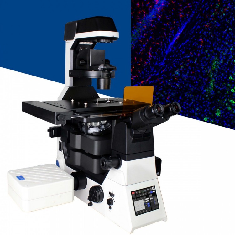
| Accessories for Microscopes | NIB1000 - AT Research-grade Inverted Microscope, Electric Type | |
| Optical System | NIS60 Infinite Range Optical System | |
| Eyepiece (Field of View) | 10x, adjustable diopter -5 to +5 | |
| Observing Tube | 10 - 40 degrees tilt, lens distance 47 - 78mm | |
| NIS60 Objective Lens | 4x Plan Apochromatic Objective, NA = 0.16, WD = 17.00mm (optional), Cover Slip Correction: 0.17mm 10x Plane Field Compensating Objective, NA = 0.45, WD = 4.00mm, Cover Glass Correction: 0.17mm 20x Plane-Apochromatic Objective, NA = 0.75, WD = 1.10mm, Cover Glass Correction: 0.17mm 40× Plan Apochromatic Objective, NA = 0.95, WD = 0.19 - 0.21mm, Cover Glass Correction: 0.17mm 60× Plan Apochromatic Objective, NA = 1.42, WD = 0.25mm, Cover Glass Correction: 0.17mm 100× Plane Achromatic Objective, NA = 1.45, WD = 0.13mm, Cover Glass Correction: 0.17mm (optional) 100× Plan Apochromatic Objective, NA = 1.49, WD = 0.09 - 0.16mm Cover glass correction: 0.13 - 0.19mm (23°C) / 0.14 - 0.20mm (37°C) | |
| Objective Converter | Electrical 6-Pin Converter (DIC Expansion Slot) | |
| Electrical six-hole converter (DIC expansion slot), with anti-focus drift module; (optional) | ||
| Converging lens | 7-hole electric rotary magnifying lens | |
| Lighting System | Transmitted C罗拉 lighting, LED lighting; | |
| Fallover Lighting: Wide Field 4 Band LED Lighting 6-hole Electric Fluorescent Turntable (B, G, U standard); Electric Fluorescent Aperture. | ||
| Mid-ratio switch | Manual 1×, 1.5× | |
| Body port | Split ratio: Eye 100%, Left Port 100%, Right Port 100%, Eye 20%/Right Port 80% | |
| Platform | Electromagnetic Control (Grating Type): Stroke Range 130mm × 100mm (Tabletop 445mm × 300mm)zuiHigh speed: 25mm/s; positioning accuracy: 0.1μm; repeatability accuracy: 0.5μm, equipped with universal holder (compatible with 35mm - 65mm petri dishes and sections), optional orifice plate holder | |
| Focusing System | Electric Z-axis, stroke 10mmzuiStep resolution 0.02μm, repeatability 0.1μm 3 Focus Adjustment Knobs (2μm/cycle, 40μm/cycle, 200μm/cycle) | |
| DIC plug-in | 10X, 20X, 40X, 60X, 100X magnification inserts, compatible with converter slots (optional) | |
| Software | Software: Nomis Pro X - C with Confocal Scanning Function, Multidimensional Imaging, and Channel-Specific Preset Features; equipped with wide-field and confocal imaging capabilities, the software features a complete control module for hardware, built-in image basic filtering, and supports multiple image output formats. | |
| Camera | Nexcam - T12 | |
| Enhanced features | Living Cell Culture System SR - SIM Ultra-Resolution Module Double-layer Fluorescent Tall Attachment | |
The application fields of the NCF1000 laser confocal microscope mainly include the following aspects:
◆Biological Research
High-resolution imaging of the internal structure of live cells or tissues
3D Image Reconstruction and Multichannel Fluorescence Analysis
Real-time dynamic observation and image capture
Scientific Research & Clinical Diagnosis
Visualization of organelle-level microstructure
Histopathology Sample Analysis
Mechanism of drug action verification and localization of traditional Chinese medicine ingredients
◆ Materials Science
Support for nanoscale characterization of new materials (such as coatings, films)
Compatibility metal/non-metal hetero-surface detection
This equipment belongs to the mid-to-high-end product line among domestic instruments, featuring high sensitivity and rapid imaging capabilities, and has been verified by practical applications in universities and research institutions.
